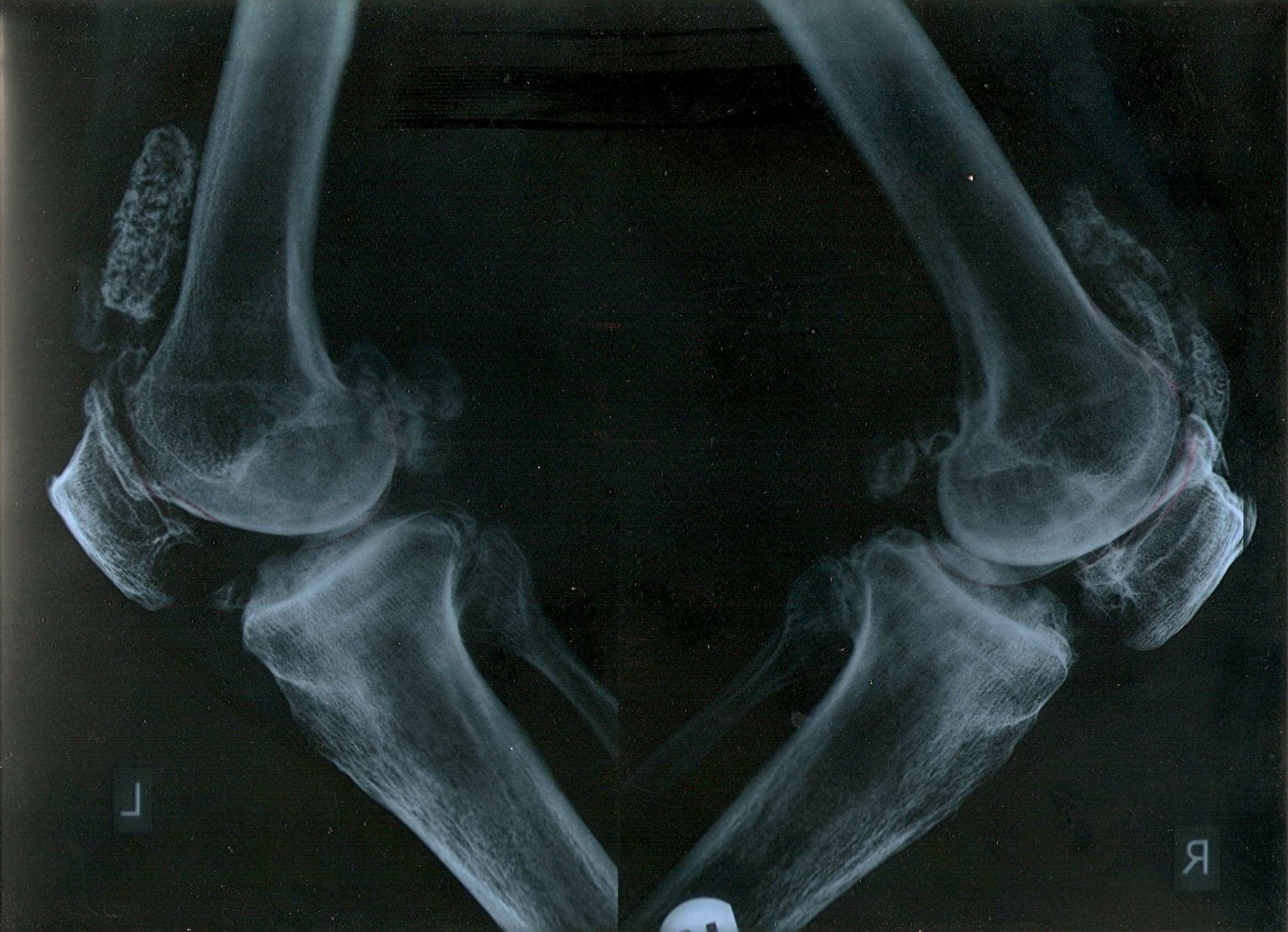
Patient
no 1
The radiographs of both the knees
below belong to a gentleman from another State in India (name and
location withheld). He was the relative of a doctor who regularly
refers to us. The doctor wished, an assessment be done to
check if surgery can be avoided, as total knee replacement had been
advised
The patient had pain every day
for the last 12 years.
He had varus and flexion
deformity in both his knees since the last several
years.
He ambled in complaining
that even walking was a problem now.
However, the left knee did not respond and
it was decided to explore the hip and the spine to examine if the
symptoms on the left knee could be changed, once again using the
Mckenzie assessment for the lumbar spine.
A
rapid reversal was encountered in his
symptoms and deformity, giving significant change to his baseline
gait, with repeated movements to the spine, thus giving us a
directional preference to treat the symptoms in his knee with
movements to the spine.The patient was naturally happy and went
home happily with a home programme designed for
him.
The patient was followed up over a
year, telephonically, and he remained better, doing his self
mangement.
We learned an important lesson
from this patient. Every patient deserves a chance for recovery,
despite how many years his symptoms are present and the deformities
he may have. The human body can be
unpredictable.
The patient had been with the
flexion and varus deformity for the last several years, but it
needed just a few movements in the right direction at the right
joints for the patient to be relieved of
it.
Patient no
2:
This is one of our overseas patient we
treated in the short time he was here. The patient (Name withheld)
was seeking treatment for left shoulder pain, and was
referrred to us by a doctor who trusted the McKenzie system. This
patient had been advised surgery for the shoulder at a tertiary
care hospital, and advised that for his condition, the results
cannot be guaranteed post surgery.
His
MRI reported a complete tear of the subscapularis tendon, a type 2
SLAP tear, hypertrophy and cystic changes in the lesser tuberosity,
soft tissue thickening with synovitis in the rotator cuff interval,
tendinosis in the infraspinatus and supraspinatus with fatty
atrophic changes in the infraspinatus.
The history suggested a shoulder disorder
and the Mckenzie extremity assessement was done on the
patient.
extensive assessment only showed us that
the shoulder was symptomatic on loading, but there was no clear
mechanical diagnosis emerging, nor was there any pattern to
classify the patient to a shoulder disorder. At the next session,
the cervical spine was assessed keeping the baselines of the
shoulder. We were able to establish during the mechanical
assessment that the positions and movements of the cervical spine
affected the patients concordant pain in the shoulder. The
management was for the cervical spine and the patient got better in
3-4 sessions symptomatically and functionally.
We followed the patient telephonically
after 1 month and after more than a year. The patient had not
sought any other treatment and continued to be without symptoms,
and continued to use his shoulder normally.
This case has now been published
in an international publication.
A. Menon, S. May. Shoulder pain:
Differential diagnosis with mechanical diagnosis and therapy
extremity assessment. A case report. Manual Therapy 18 (2013)
354e357
Some research
evidences -
It is important for better outcomes to treatment and the
prognosis, that an accurate differentialtion is done between
shoulder and cervical disorders causing shoulder pain (Mannifold and McCann,
1999).
The clinical tests for making a pathoanatomic diagnosis to the
shouldder do not have good levels of reliability (May et al., 2010) or validity
(Dinnes et al., 2003;
Mircovic et al., 2005; Dessaur and Magarey, 2008; Hegedus et al.,
2008; Hughes et al., 2008).
Patient no
3
A 65 year old lady was brought to
us for favor of treatment for her knee pain by her daughter. She
had knee pain since 3-4 months prior to visiting this clinic. The
patient was once again advised a Total Knee Replacement, based on
the radiographic findings and failure to reduce the symptoms and
attain normal functions with other conservative
treatment.
The history revealed that the knee
pain was a sudden onset, and this was the first episode of knee
pain. The patient had been fully functional with normal Indian
squatting, cross legged floor sitting, ascending and descending
stairs independently with no pain whatso-ever, till the
sudden onset of pain 3-4 months prior.
The patient's knee, and hip
was explored for possible source of pain. A mechanical assessment
using the McKenzie lumbar assessment form was then done in order to
examine if the movements, positions, postures of the lumbar spine
affected the symptoms of the knee.
The assessment gave us a
directional preference which not only abolished the knee pain
rapidly, but also restored the patient to normal functions. The
patient went through 4-5 sittings to become fully restored to
normal functions.
Post treatment this patient was followed
up at 3 months and 6 months. She had continued to remain pain free,
fully restored to functions and had not sought any other treatment
elsewhere.
This case was presented during the
Private Practisioners conference in Mumbai
in
2007.